
dicom-viewer
3d-visualization
radiology-processing
nuclear-medicine
Medical imaging software for viewing and processing DICOM images, 2D, 3D, and 4D visualization with advanced radiology and nuclear medicine tools, runs natively on Mac OS X.
What is OsiriX?
OsiriX is a feature-rich, open-source medical imaging software designed specifically for viewing and processing DICOM images. It provides powerful 2D, 3D, and 4D visualization capabilities along with a comprehensive set of processing tools for radiology and nuclear medicine applications.
Some key features of OsiriX include:
- Native support for DICOM images including CT, MRI, PET, SPECT, X-ray Angiography, Ultrasound, and more
- 2D multiplanar reconstruction with scroll, pan, zoom, cine loop, and more
- 3D volume rendering with MIP, MinIP, Avg, SSD, VR render modes
- 4D time-resolved imaging for dynamic studies
- Advanced image processing filters, ROI measurements, image fusion, and image registration
- Structured reporting, key images, and annotations
- Plugin architecture for extending functionality
- DICOM networking, printing, importing/exporting, CD burning
A key advantage of OsiriX is that it is designed specifically for Mac OS X to leverage its speed, graphics capabilities, and stability. This makes it a robust choice for day-to-day use in reading and manipulating large DICOM study sets.
OsiriX Features
Features
- 2D, 3D and 4D DICOM image visualization
- Multiplanar reconstruction
- Volume rendering
- Image fusion
- ROI tools
- DICOM networking
- Plugin architecture
Pros
Free and open source
Native Mac OS X application
Wide range of visualization and processing tools
Supports many DICOM formats
Active user and developer community
Cons
Limited support for some advanced visualization techniques
Steep learning curve
Mac OS X only
Official Links
The Best OsiriX Alternatives
Top
Medical
and
Radiology Software
and other similar apps like OsiriX
Horos
Horos is an open-source, cross-platform medical image viewer software designed specifically for viewing and analyzing DICOM and other medical images. Originally based on the OsiriX image viewer, Horos has been completely rewritten with an improved user interface and additional features.Key features of Horos include:Intuitive interface for browsing complex DICOM studies…

Weasis
Weasis is an open source platform for viewing and analysing digital medical images. It is implemented in Java and can run on most operating systems (Windows, Mac OS, Linux). Weasis supports viewing and processing DICOM images as well as JPEG, PNG and other common image formats.Some key features of Weasis…

3D Slicer
3D Slicer is a free, open source platform for medical image computing. It provides visualization and image analysis tools for various medical imaging modalities including MRI, CT, US. Its main features include:Multi-modal 2D and 3D visualization of medical image dataImage segmentation using both manual and semi-automated techniquesRegistration of images from…

3DimViewer
3DimViewer is a versatile, free, open-source program for interactively viewing 3D models from a variety of fields. It supports common 3D file formats including OBJ, OFF, PLY, STL, and more, allowing users to easily view 3D data.Key features of 3DimViewer include:Intuitive user interface for rotating, panning, zooming, and manipulating 3D…

AMIDE
AMIDE (Amide’s a Medical Image Data Examiner) is a free, open source software application for viewing, analyzing, and registering volumetric medical imaging data sets. It supports DICOM and ANALYZE image formats.Some of the key features of AMIDE include:Loading and visualization of DICOM, ECAT, ANALYZE, and raw binary medical imagesTools for…

DicomWorks
DicomWorks is a free, open source DICOM viewer, editor, converter, and toolset software for Windows, Mac OS, and Linux operating systems. It enables medical professionals, researchers, and patients to easily view, edit, annotate, process, and convert DICOM medical images, datasets, and documents.Key features include multi-frame stacking and scrolling, basic viewing…

MicroDicom
MicroDicom is a free, open-source DICOM viewer, editor and converter for Windows. It is designed to be a lightweight and easy-to-use application to view, edit and convert DICOM medical images and documents.Some of the key features of MicroDicom include:DICOM image viewer with pan, zoom and window/levelDICOM editor to update tags…

Materialise Mimics
Materialise Mimics is a medical imaging software platform developed by Materialise for working with medical image data from CT and MRI scans. It provides sophisticated tools for 3D visualization, image segmentation, analyzing measurements, and creating 3D models from stacks of medical images.Some key features and capabilities of Materialise Mimics include:Advanced…

DICOMan
DICOMan is an open-source toolkit for working with medical images stored in the Digital Imaging and Communications in Medicine (DICOM) format. It can be used to organize, view, process, and analyze DICOM images and related data.Some key features of DICOMan include:DICOM file management — Import, export, query, and organize a…

Vesalius3D
Vesalius3D is an open-source software application designed specifically for creating detailed and accurate 3D visual models of human anatomy. It incorporates powerful image processing and visualization capabilities that allow users to import medical imaging datasets such as CT or MRI scans and process the scan data to reconstruct anatomical structures…

3D-DOCTOR
3D-DOCTOR is a powerful 3D modeling and image processing software designed specifically for MRI and CT scan analysis. It enables medical professionals to easily reconstruct 3D models from scan images and accurately measure and visualize anatomical structures.Some key features of 3D-DOCTOR include:Import DICOM files directly from MRI or CT scannersSegment…

Navegatium
Navegatium is an open-source web browser that prioritizes user privacy and security. It is built on Chromium, but unlike Chrome, Navegatium blocks online ads and trackers by default to prevent companies from collecting data on your browsing habits.Some key features of Navegatium include:Blocks ads, trackers, cryptocurrency mining, and fingerprinting techniques…

Starviewer
Starviewer is an open source DICOM viewer software designed for medical imaging. It can open and visualize DICOM images and supports features like 2D viewer, 3D volume rendering, 4D animation, multiplanar reconstruction, fusion visualization, cine loop, image filters and measurements.As an open source software, Starviewer is free to download and…

Ginkgo CADx
Ginkgo CADx is an open-source DICOM viewer and diagnostic tool designed specifically for medical imaging and analysis. It provides a comprehensive set of tools for viewing, processing, analyzing, and exporting medical images in the DICOM standard.Some of the key features of Ginkgo CADx include:Multi-modality DICOM viewing for modalities like CT,…

InVesalius 3
InVesalius is a free open-source software for medical imaging 3D reconstruction from DICOM files. It was developed at the Center for Information Technology Renato Archer (CTI) in Brazil.InVesalius allows users to import tomographic images in DICOM format from CT or MRI scanners. It can generate 3D surface models from imported…

Miele-LXIV
Miele-LXIV is a next-generation business intelligence and analytics platform designed to help companies make data-driven decisions. It provides tools for data preparation, visualization, augmented analysis, and sharing insights across the organization.Some key features of Miele-LXIV include:Intuitive drag-and-drop interface for building dashboards with advanced charts, tables, KPIs and moreAugmented analysis using…

Hipax Diagnostic Workstation
Hipax Diagnostic Workstation is an advanced medical imaging software solution used by radiologists, clinicians, and imaging technicians for viewing, post-processing, analyzing, and reporting medical images from modalities like X-ray, CT, MRI, PET, SPECT, and more.It provides a comprehensive set of tools for multi-modality viewing, 3D MIP/MPR reconstructions, image fusion, quantitative…
DICOMscope
DICOMscope is a powerful, multi-platform DICOM viewer and editor. As an open-source application, it provides a free alternative to proprietary DICOM software.It supports viewing and processing images from modalities like CT, MRI, PET, ultrasound, and x-ray. Images can be rendered in both 2D and 3D, allowing radiologists and technicians to…

GIMIAS
GIMIAS (Graphical Interface for Medical Image Analysis and Simulation) is an open-source software platform designed for biomedical research and clinical evaluation. It provides a graphical workflow editor and library of processing and simulation tools that allow users to create and execute complex pipelines for medical image computing and analysis.Some key…

Dicompyler
dicompyler is an open source software platform built on top of the DICOM standard for radiation therapy research. It is designed to help researchers view, analyze, process, and convert DICOM RT data sets.Some of the key features of dicompyler include:Importing and exporting DICOM RT objects like images, structure sets, plans,…

XMedCon
xMedCon is a free, open-source software application designed specifically for medical image format conversion. It supports converting between DICOM (.dcm), NIfTI (.nii), Analyze (.hdr/.img), NRRD (.nrrd), PNG (.png), JPEG (.jpg), TIFF (.tif), and other common medical imaging formats.Some key features of xMedCon include:Intuitive graphical user interface for easy batch converting…

12 подробностей о OsiriX HD
1. OsiriX HD is capable of displaying images from all imaging modalities (ultrasound, CT scanner, MRI, PET, etc.) in their native standard DICOM format used by the medical/scientific industry.
2. OsiriX HD is a fully DICOM-compliant listener that can receive images from any DICOM imaging device through WiFi/3G networks.
3. «OsiriX HD» is a DICOM software for iOS: DICOM is the digital standard for storing and transferring medical images.
4. The convenience of being able to access DICOM images remotely added to the fact that images can be previewed on the iPhone/iPad screen in a very effective and convenient way offers a new perspective for mobile teleradiology.
5. OsiriX HD provides fast interactive image manipulation tools such as zoom, pan, cine and contrast adjustment of images through the multipoint touch screen interface.
6. OsiriX HD is a full DICOM image viewer for iOS (DICOM Files & DICOM Network protocol support).
7. This iOS application can also be used as a helper application for other iOS applications: it allows to read DICOM datasets received by emails or in Safari, or to visualize DICOM datasets stored in DropBox folder, for example.
8. Initial experience with a handheld device digital imaging and communications in medicine viewer: OsiriX mobile on the iPhone.
9. This iOS app is designed to work seamlessly with any DICOM compatible software, including PACS, medical workstations, acquisition modalities.
10. OsiriX HD allows downloading and manipulating series of images directly on your iOS device.
11. It supports these DICOM Network protocols: C-STORE SCP, C-MOVE SCU, C-FIND SCU, C-GET SCU, WADO, DICOMweb.
Если вам нравится OsiriX HD, вам понравятся Medical такие приложения, как; РТ ДокторОнлайн; Умный мониторинг здоровья; ТРЭК 2022; BCT Taxonomy; BigMagnify; AMBOSS Medical Knowledge; Master Tung Acupoint; Vet Manual : Animal Diseases; АнтиПаника; Доказательная кардиология; Анатомия — 3D Атлас; Sanford Guide — Hepatitis; Healing – Self Love Hypnosis; Календарь Менструации; ReSound Tinnitus Relief; Kwarantanna domowa; Apteka.ru – заказ лекарств; Elsevier CDI Reference; ТИННИТУС, БЕЛЫЙ ШУМ, СОН, ХРАП; Elsevier Clinical Pharmacology;
Программа для просмотра МРТ, КТ и рентгеновских снимков
После прохождения МРТ или КТ исследования у пациента часто возникает желание собственноручно посмотреть какие изменения выявил у него врач. Файлы исследования обычно имеют формат DICOM (*.dcm). Для того, чтобы открыть файлы с данным разрешением требуется специальный просмоторщик из группы программ для радиологии. Группа программ для просмотра КТ, МРТ и рентгенограмм называется на английском DICOM viewer, а по-русски программа для просмотра DICOM файлов. Для того, чтобы найти одну программу из данной группы достаточно ввести в поисковик «программа для просмотра DICOM файлов». Лучше всего в конце запроса указать вашу операционную систему (Windows XP, Windows Vista, Windows 7,8,10 или Mac OS Leon, Leopard, Yosemite, Capitan). Для Mac OS наиболее удачные программы — это OsiriX и Horos. Большой плюс этих программ является то, что их можно использовать и на бесплатной основе, что для единичного пользования программы пациентом немаловажно. Данные программы также используют ведущие радиологи в мире.

Программа для просмотра КТ снимков.
Для того, чтобы просмотреть файлы с диска вам потребуется сохранить (скопировать) информацию с диска на жесткий диск компьютера, а после открыть данный архив в программе для просмотра снимков. Ваши снимки возможно визуализировать в аксиальной, сагиттальной и фронтальной проекции. Если при МРТ исследовании врач радиолог должен настроить аппарат, чтобы получить изображения во всех трех плоскостях, то при компьютерной томографии ваши изображения будут получены в аксиальной проекции. При использовании программы вы можете собственноручно переделать КТ-сканы из аксиального сечения в сагиттальное или фронтальное сечение. При помощи данных программ вам удастся также получить 3D изображение. Языком лучевой диагностики — это называется создание мультипланорной реконструкции.
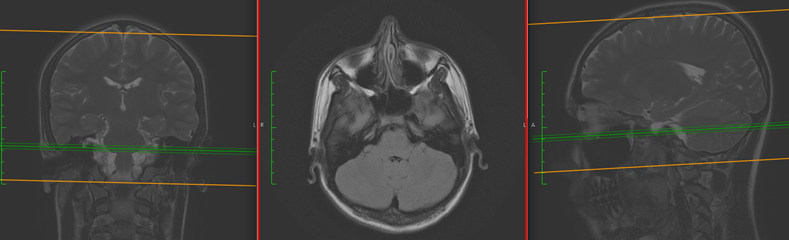
МРТ головного мозга у пациента 13 лет по поводу головных болей. Представлены три проекции. Слева — фронтальная проекции. Посередине — аксиальная проекция. Справа — сагиттальная проекция
Программа для просмотра рентгеновских снимков с диска.
Многие производители программ предлагают пробный 30 дневный период. Для того, чтобы посмотреть пациенту единожды свой снимок этого достаточно, а для работы радиологу нет. Для загрузки одного из таких приложений перейдите по следующей ссылке:
http://www.radiantviewer.com/download.php

Предложенная программа называется Radiantviewer. Оптимально работает на следующих Windows XP (service pack 3), Windows Vista, Windows 7, Windows 8 и 8,1, Windows 10. Данная программа очень проста в использовании так, как максимально интуитивна. Большим плюсом этой программы, что она переведена на русский язык от разработчика. Небольшой бонус простоты использования заключается в том, что пользователю не нужно загружать файлы с дисков на компьютер, а программа сделает это за вас, открыв автоматически ваши исследования с диска. Данная программа не потребует от вас дополнительных приложений таких, как JAVA или NET, что значительно облегчает процесс просмотра DICOM — файлов.
Данная программа поддерживает DICOM файлы следующих исследований:
1) цифровая рентгенография и маммография.
2) МРТ — магнитно-резонансная томография.
3) КТ — компьютерная томография.
4) УЗИ — ультразвуковое исследование.
5) ЦА — цифровая ангиография.
6) ПЭТ/КТ — позитронно-эмисионная томография.
Просмотр снимков мрт.
Программа RadiAnt позволяет работать с одинаковой хорошей скоростью и на компьютерах с оперативной памятью 512 мегабайт, а также на компьютерах с оперативной памятью 4 гигабайт и выше. Конечно, на компьютерах более мощных у пользователя больше возможностей для использования всего потенциала данной программы.
В программе пользователь может выполнить следующие действия:
1) Изменение яркости и контрастности.
2) Увеличение или уменьшение объекта.
3) При оценке КТ возможность выбора окна визуализации (легочное, мягко-тканное, костное).
4) Поворот, разворот, зеркальное отражение сканов.
5) Произвести замеры длины, толщины, ширины, объема.
6) Измерение плотности тканей в единицах Хаунсфильда при КТ исследовании.
При использовании данной программы у вас есть возможность сохранить DICOM изображение, как видео в формате WMV или изображение в формате JPEG. Также вы можете скопировать изображение в буфер обмена, а далее использовать изображении в презентации или заключении, что удобно при подготовке доклада с презентацией и для более информативного заключения с изображением.
Программа для просмотра мрт снимков скачать
Бесплатные программы для Mac OS являются Osirix и Horos. Данные программы возможно скачать по следующим ссылкам:
OsiriX — http://www.osirix-viewer.com/Downloads.html
Horos — http://www.horosproject.org/download/
Для того, чтобы скачать Horos вам нужно будет заполнить следующий формуляр:
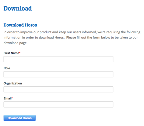
First Name — ваше имя. Role — ваша профессия. Organization — ваше место работы. Email — ваш электронный адрес. Буквально через несколько секунд вам на почту должно прийти ссылка на скачивания просмоторщика в dmg — формате. Далее устанавливаете программу, как обычно, устанавливаете программу в Mac OS.
Программа для просмотра мрт снимков.
На мой взгляд лучшая программа для Mac OS является Horos. Да эти программы (Osirix и Horos) обе бесплатные, но Horos не требует перейти на платный режим Osirix MD. При просмотре не будет выявляться красным по черному «Not for medical usage», что в переводе не для медицинского использования. Вы просто пользуетесь замечательной бесплатной программой Horos, которая вас беспокоит лишь, когда нужно загрузить обновление. В данной программе возможно открывать DICOM файлы следующих изображений: цифровая рентгенография и маммография, МРТ — магнитно-резонансная томографии, КТ — компьютерная томографии, УЗИ — ультразвуковое исследование, ЦА — цифровая ангиография, ПЭТ/КТ — позитронно-эмисионная томография.
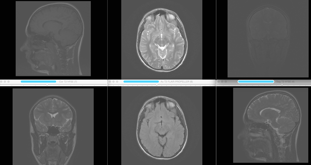
При исследовании в трех проекция легче выявить патологию. Многие радиологи используют сразу шесть окон для оценки патологии. Особенно это характерно для МРТ исследований, когда требуется оценка патологии в разных режимах T1, T2, FLAIR, STIR, DWI, T1 + contrast.
Программы просмотра рентгеновских снимков
Раньше рентгенограммы печатались на пленку, а сейчас все снимки являются цифровыми. Для того, чтобы просмотреть данные снимки требуются те же программы, которые используются при просмотре МРТ, КТ и ПЭТ/КТ снимки. Это является очень удобным в связи с тем, что у пользователя есть возможность сравнить изменения на КТ, МРТ и рентгенограмме в одном окне, к тому же это очень информативно.

Хотелось бы привести пример использования программы.
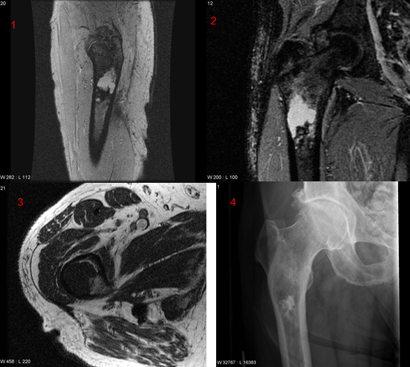
Выявлено образование в проксимальной части бедренной кости на рентгенограмме. Далее пациенту было назначено МРТ бедренной кости. Для того, чтобы понять какие анатомическо-структурные изменения выявлены у пациента обязательно нужно сравнить с рентгенограммой.
Программа для просмотра мрт снимков.
На данных четырех изображениях представлен один и тот же пациент. Первое изображение МРТ T2 fatsat в сагиттальной проекции. Второе изображение МРТ STIR в корональной проекции. Третье изображение МРТ Т1 изображение. Четвертое изображение — рентгенограмма. На разных последовательностях очаг характеризуется по-разному, что дает возможность охарактеризовать процесс более детально.
Программа для просмотра КТ снимков.
Программы для просмотра КТ снимков имеют следующие действия:
1) Изменение яркости и контрастности.
2) Увеличение или уменьшение объекта (ZOOM).
3) Выбор окна визуализации (легочное, мягко-тканное, костное).
4) Поворот, разворот, зеркальное отражение сканов.
5) Замер длины, толщины, ширины, объема.
6) Вычисление плотности тканей в единицах Хаунсфильда.
Также не маловажным критерием для программы является 3D трансформация изображения, что особенно важно при патологии сосудистой системы и при травмах.
Компьютерная томограмма 3d — реконструкция. На современных программах есть возможность выделить отдельно систему органов. В данном случае отдельно выделена сердечно-сосудистая система. У данного пациента поражена брюшная часть аорты (указана стрелкой). Болезнь Такаясу.
У данного пациента назо-орбито-этмоидальный перелом. 3d помогает в подборе хирургической тактики.
Другие
статьи из раздела «Общие вопросы МРТ»
Preview
WindowsDen the one-stop for Medical Pc apps presents you OsiriX HD by Pixmeo SARL — OsiriX HD is a full DICOM image viewer for iOS (DICOM Files & DICOM Network protocol support).
AWARDS
— Aunt Minnie’s 2013 — «Best Radiology Mobile App”
— Mobie Awards 2009: Winner — Best Medical App
— RSNA 2008: Excellence in Design
— RSNA 2008: Certificate of Merit
«OsiriX HD» is a DICOM software for iOS: DICOM is the digital standard for storing and transferring medical images. OsiriX HD allows downloading and manipulating series of images directly on your iOS device. OsiriX HD is capable of displaying images from all imaging modalities (ultrasound, CT scanner, MRI, PET, etc. ) in their native standard DICOM format used by the medical/scientific industry. OsiriX HD is a fully DICOM-compliant listener that can receive images from any DICOM imaging device through WiFi/3G networks.. We hope you enjoyed learning about OsiriX HD. Download it today for £48.99. It’s only 28.84 MB. Follow our tutorials below to get OsiriX HD version 5.5.6 working on Windows 10 and 11.
GET OsiriX HD for PC
After checking the 💻 Windows AppStore, we found the following OsiriX HD apps (Available for Direct download):
OR
Alternatively, download OsiriX HD APK for PC (Emulator) below:
Download OsiriX HD APK for use on PC
| Download | Developer | Rating | Reviews |
|---|---|---|---|
|
IDV — IMAIOS DICOM Viewer
Download Apk for PC ↲ |
IMAIOS SAS | 3.9 | 447 |
|
IDV — IMAIOS DICOM Viewer
GET ↲ |
IMAIOS SAS | 3.9 | 447 |
|
mRay
GET ↲ |
mbits imaging GmbH | 3.3 | 181 |
|
MRI Essentials
GET ↲ |
mr-verlag | 4.2 | 74 |
|
DroidRender — 3D DICOM viewer
GET ↲ |
Startm | 4.5 | 638 |
|
e-Anatomy
GET ↲ |
IMAIOS SAS | 4.2 | 3,185 |
Follow Tutorial below to use OsiriX HD APK on PC:
- Download an Android App emulator.
An emulator imitates/ emulates an android device on your computer, making it easy to install and run android apps from the comfort of your PC. We’ve listed the best below:- Nox »
- Bluestacks »
- Install the Bluestacks.exe or Nox.exe Software emulator on your Windows PC.
- Run OsiriX HD on PC:
- Once installed, open the Emulator app, type OsiriX HD in the search bar and search.
- You will immediately see OsiriX HD. Open it and click Install. The app will start Installing.
- To run the OsiriX HD app on PC, open Emulator » «All Apps» » OsiriX HD.
Get OsiriX HD on Apple Mac
5.8
Mavericks 10.9 compatible
5.7.1
Minor GUI corrections, and optimizations
5.7
- OsiriX MD is FDA / CE certified as Medical Device Class II
- DICOM: Support for Breast Tomosynthesis Image Storage
- DICOM: Support UTF8 string for ROI description box, instead of ASCII 127 only
- DICOM: Support for SUV calculation on PET series with decayCorrection NONE and ADMIN
- DICOM: Support for EnhancedUSVolumeStorage
- DICOM: Support for EncapsulatedCDAStorage
- DICOM: Support for EstimatedRadiographicMagnificationFactor
- DICOM: Merge Studies: option to modify DICOM files
- DICOM: Query Retrieve window: Option to have smaller refresh intervals
- DICOM: WADO Retrieve: Start retrieving before C-FIND ends
- DICOM: Multiple associations during DICOM STORE-SCU (DICOM Send)
- PACS On-Demand: Automatically retrieve partial studies
- PACS On-Demand: Support for next/previous patient
- PACS On-Demand: Support for last name and first name searches
- ROI: Anti-aliased ROI text rendering
- ROI: Display radii for Circle ROIs
- ROI: Option to display ROI info, only when mouse is over
- ROI: 90, 45, 0 lines drawing when pressing shift key
- ROI: Support alpha pixel for tBrush selection
- ROI: Compute skewness / kurtosis
- ROI: Display units, if available
- ROI: Support ROI mean, min, max calculation for RGB images
- 3D Curved-MPR: Display transverse lines with real size according to transverse view zoom level
- 3D Curved-MPR: Option to enter thickness manually (similar to MPR)
- 3D Curved-MPR: Export window: option to enter the slice interval, manually
- Database: Add ‘Day Before Yesterday’ option for Smart Albums
- Database: Display both ages: actual and at time of study
- Database: Faster Database delete process
- Database: Display progress bar for WADO download
- 2D viewer: Next/Previous Patient button: load workspace state if available
- 2D viewer: Display if VOI LUT is applied or available
- 2D viewer: Display the thickness in mm
- 2D Viewer: Always order the windows from most recent study to oldest study
- 2D Viewer: Display study color / number, based on all available studies, not only on displayed studies
- 2D Viewer: Display overflow lines if raw data is not completely displayed in the view
- Hanging Protocols: Support for comparatives
- Preferences: Centralized Defaults Preferences
- Preferences: Limit auto-comments to study or series level
- Preferences: Activate checkbox for DICOM nodes
- Preferences: Erase entire DB at each startup / quit
- Preferences: Completely ignore CD/DVD
- WebPortal: For administrators: delete studies and consult network logs
- WebPortal: Other studies for a patient: Sorted by Date
- WebPortal: Flash sequence: double-click on the image to auto-play / stop
- WebPortal: Flash sequence: Play movie at defined frameRate WebPortal: Recent patients list
- WebPortal: Faster algorithm for displaying the users list allowed to view a study
- WebPortal: Progress indicator when uploading a file